- Startseite
- Forschung
- Neonatologie
- Translationale Neonatologie
- Plazentare Störungen
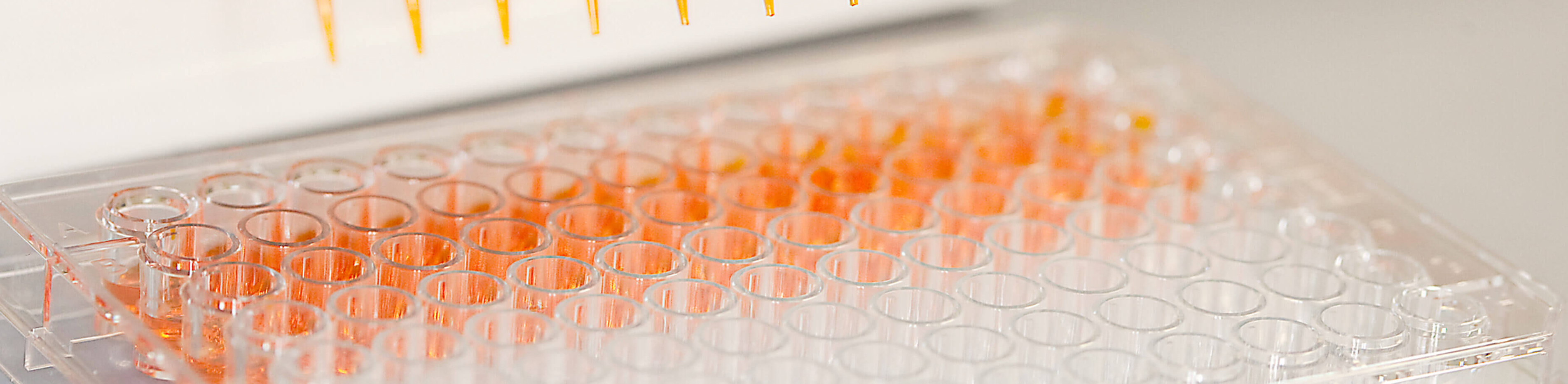
- Placental dysfunction
Our group is focussing on the influence of the perinatal environment on the infant’s development. In particular, we are interested in the function of the placenta due to its essential role in providing supply of the fetus with nutrients, oxygen and hormones in utero. Dysfunction of the placenta is known to result in changes of perinatal programming and hence, in long term consequences for the child.
Placental dysfunction does originate from several factors. However, in our lab we are focusing on placental disturbances caused by maternal obesity and by an inflammatory environment. Besides revealing the molecular mechanisms that lead to a disturbed placental function, it is extremely important to us finding specific and adequate interventions to prevent those negative impacts. To do so we are working with animal and cell culture models, but also use human placentas for our research. In this context, the translational aspect is very important for us.
Our long term goal is to develop new diagnostic and therapeutic options to prevent negative effects on the perinatal supply which in turn might cause problems in later life.
The Team
Dr. Eva-Maria Turnwald
Maria Wohlfarth
* these authors contribute equally to this work
Kretschmer T, Turnwald EM, Janoschek R, Zentis P, Bae-Gartz I, Beers T, Handwerk M, Wohlfarth M, Ghilav M, Bloch W, Hucklenbruch-Rother E, Dötsch J, Appel S (2020). Maternal high fat diet-induced obesity affects trophoblast differentiation and placental function in mice. Biol Reprod. 103:1260-1274.
Pichl T, Keller T, Hünseler C, Roth B, Janoschek R, Appel S, Hucklenbruch-Rother E (2020). Ketamine effects on neurogenesis, extracellular matrix homeostasis and proliferation in hypoxia-exposed murine hippocampal neurons. Biomed Rep. 13:23
Bae-Gartz I*, Kasper P*, Großmann N, Breuer S, Janoschek R, Kretschmer T, Appel S, Schmitz L, Vohlen C, Quaas A, Schweiger MR, Grimm C, Fischer A, Ferrrari N, Graf C, Frese C, Lang S, Demir M, Schramm C, Fink G, Goeser T, Dötsch J, Hucklenbruch-Rother E (2020). Maternal exercise conveys protection against NAFLD in the offspring via hepatic metabolic programming. Sci Rep 10:15424.
Kretschmer T, Schulze-Edinghausen M, Janoschek R, Handwerk M, Wohlfarth M, Hucklenbruch-Rother, Dötsch J and Appel S (2020). Effect of Maternal Obesity in Mice on IL-6 Levels and Placental Endothelial Cell Homeostasis. Nutrients 12: pii: E296.
Nüsken E, Turnwald EM, Fink G, Voggel J, Yosy C, Kretschmer T, Handwerk M, Wohlfarth M, Weber LT, Hucklenbruch-Rother E, Dötsch J, Nüsken KD, Appel S (2019). Maternal high fat diet and in-utero metformin exposure significantly impact upon the fetal renal proteome of male mice. J. Clin. Med, J. Clin. Med. 8: pii: E663.
Bae-Gartz I, Janoschek R, Breuer S, Schmitz L, Hoffmann T, Ferrari N, Branik L, Oberthuer A, Kloppe CS, Appel S, Vohlen C, Dötsch J, Hucklenbruch-Rother E (2019). Maternal obesity alters neurotrophin-associated MAPK signaling in the hypothalamus of male mouse offspring. Front Neurosci. 13:962.
Appel S, Grothe J, Storck S, Janoschek R, Bae-Gartz I, Wohlfarth M, Handwerk M, Hucklenbruch-Rother E, Gellhaus A, and Dötsch J (2019). A potential role for GSK3beta in glucose-driven intrauterine catch-up growth in maternal obesity. Endocrinology. 160:377-386.
Schmitz L, Kuglin R, Bae-Gartz I, Janoschek R, Appel S, Mesaros A, Jakovcevski I, Vohlen C, Handwerk M, Ensenauer R, Dötsch J, Hucklenbruch-Rother E (2018). Hippocampal insulin resistance links maternal obesity with impaired neuronal plasticity in adult offspring. Psychoneuroendocrinology 89:46-52.
Hucklenbruch-Rother E, Appel S, Bae-Gartz I, Schmitz L, Janoschek R, Dötsch J (2018). Perinatale Programmierung - eine Übersicht. ZS Internstische Praxis. [Review]
Ferrari N*, Bae-Gartz I*, Bauer C, Janoschek R, Koxholt I, Mahabir E, Appel S, Alejandre Alcazar MA, Grossmann N, Vohlen C, Brockmeier K, Dötsch J, Hucklenbruch-Rother E and Graf C (2018). Exercise during pregnancy and its impact on mothers and offspring in humans and mice. J Dev Orig Health Dis. 9:63-76.
Appel S*, Schulze-Edinghausen M*, Kretschmer T*, Storck S, Janoschek R, Bae-Gartz I, Handwerk M, Wohlfarth M, Nüsken KD, Hucklenbruch-Rother E, Heykants M, Mahabir E and Dötsch J (2017). Maternal obesity attenuates predelivery inflammatory reaction in C57Bl/6N mice. J Reprod Immunol. 122:10-13.
Kasper P, Vohlen C, Dinger K, Mohr J, Hucklenbruch-Rother E, Janoschek R, Köth J, Matthes J, Appel S, Dötsch J, Angel Alejandre Alcazar M (2017). Renal Metabolic Programming is Linked to the Dynamic Regulation of a Leptin-Klf15 Axis and Akt/AMPKα Signaling in Male Offspring of Obese Dams. Endocrinology 158:3399-3415.
Bultmann-Mellin I, Dinger K, Debuschewitz C, Loewe KMA, Melcher Y, Plum MTW, Appel S, Rappl G, Willenborg S, Schauss AC, Jüngst C, Krüger M, Dressler S, Wempe F, Alcázar MAA and Sterner-Kock A (2017). Role of Ltbp-4 in alveolarization, angiogenesis and fibrosis in lungs. Am J Physiol Lung Cell Mol Physiol. 313:L687-L698.
Nüsken E, Appel S, Hucklenbruch-Rother E, Dötsch J, Nüsken KD, Alejandre Alcazar MA (2017). Umwelteinflüsse in der Schwangerschaft – Risiko für die Krankheitsdisposition im späteren Leben. Monatsschrift Kinderheilkunde 165:379–388. [Review]
Janoschek R, Bae-Gartz I, Vohlen C, Alcazar MA, Dinger K, Appel S, Dötsch J and Hucklenbruch-Rother E (2016). Dietary intervention in obese dams protects male offspring from WAT induction of TRPV4, adiposity, and hyperinsulinemia. Obesity (Silver Spring) 24:1266-1273.
Nüsken E, Herrmann Y, Wohlfarth M, Goecke T, Appel S, Schneider H, Dötsch J, Nüsken KD (2015). Strong hypoxia reduces leptin synthesis in purified primary human trophoblasts. Placenta 36:427-432.
Appel S*, Turnwald EM*, Ankerne J, Wohlfarth M, Appel J, Rother E, Janoschek R, Alejandre-Alcazar MA, Schnare M, Meißner U and Dötsch J (2015). Hypoxia mediated soluble fms-like tyrosine kinase-1 (sFlt-1) increase is not attenuated in IL-6 deficient mice. Reprod Sci. 22:735-742.
Appel S, Ankerne J, Appel J, Oberthuer A, Mallmann P, Dötsch J (2014). CNN3 regulates trophoblast invasion and is upregulated by hypoxia in BeWo cells. PLoS One 22:e103216.
Appel S*, Turnwald EM*, Alejandre Alcazar MA, Ankerne J, Rother E, Janoschek R, Wohlfarth M, Vohlen C, Schnare M, Meißner U and Dötsch J (2014). Leptin does not induce an inflammatory response in the murine placenta. Hormone and Metabolic Research 46:384-389.
Alejandre Alcazar MA, Ostreicher I, Appel S, Rother E, Vohlen C, Plank C, Dötsch J (2012). Developmental regulation of inflammatory cytokine-mediated Stat3 signaling: the missing link between intrauterine growth restriction and pulmonary dysfunction. Journal of Molecular medicine 90: 945-957.
Jensen MH, Watt J, Hodgkinson J, Gallant C, Appel S, El-Mezgueldi M, Angelini TE, Morgan KG, Lehman W and Moore JR (2012). Effects of basic calponin on the flexural mechanics and stability of F-actin. Cytoskeleton 69:49-58.
Gallant C*, Appel S*, Graceffa P, Leavis P, Lin JJ-C, Gunning PW, Schevzov G, Chaponnier C, DeGnore J, Lehman W and Morgan KG (2011). Tropomyosin variants describe distinct functional subcellular domains in differentiated vascular smooth muscle cells. Am J Physiol Cell Physiol. 300:C1356-65.
Appel S and Morgan KG (2010). Scaffolding proteins and non-proliferative functions of ERK1/2. Commun Integr Biol. 3: 354-356. [Addendum]
Appel S, Allen PG, Vetterkind S, Jin J-P and Morgan KG (2010). H3/Acidic calponin: An actin-binding protein that controls ERK1/2 activity in non-muscle cells. Mol Biol Cell. 8:1409-1422.
Zabransky S, Alejandre Alcazar M, Appel S, Bilbao N, Dötsch J, Eggermann T, Ertan AK, Ertan A, Fahlbusch F, Finken MJJ, Harder T, Haverkamp F, Hübler A, Huppertz B, Kabbur PM, Kaesmann-Kellner B, Kuschewski R, Martin DD, Nüsken E, Nüsken KD, Olbertz DM, Parikh N, Plagemann A, Plinkert PK, Reinehr T, Rochow N, Rother E, Saenger P, Schild R, Schweizer R, Straube S, Struwe E, Trollmann R, Tzschoppe A, Voigt M, Vuguin P, van de Weyer PS (2013). Caring for children born small for gestational age. Springer Healthcare. [Book]
Appel S*, Vetterkind S*, Koplin A, Maertens B, Boosen M, and Preuss U (2009). EFP1 is an ER stress-induced glycoprotein which interacts with the pro-apoptotic protein Par-4. Cell Health and Cytoskeleton. 1: 1–16.
Gangopadhyay SS*, Kengni E*, Appel S*, Gallant C, Kim HR, Leavis P, DeGnore J and Morgan KG (2009). Smooth muscle archvillin is an ERK scaffolding protein. J Biol Chem. 284: 17607-17615.
Kim HR, Appel S, Vetterkind S, Gangopadhyay S, and Morgan KG (2008). Smooth muscle signaling pathways in health and disease. J. Cell. Mol. Med. 12: 2165-2180. [Review]
Vetterkind S, Illenberger S, Kubicek J, Boosen M, Appel S, Naim HY, Scheidtmann KH and Preuss U (2005). Binding of Par-4 to the actin cytoskeleton is essential for Par-4/Dlk-mediated apoptosis. Exp. Cell Res. 305: 392-408.